
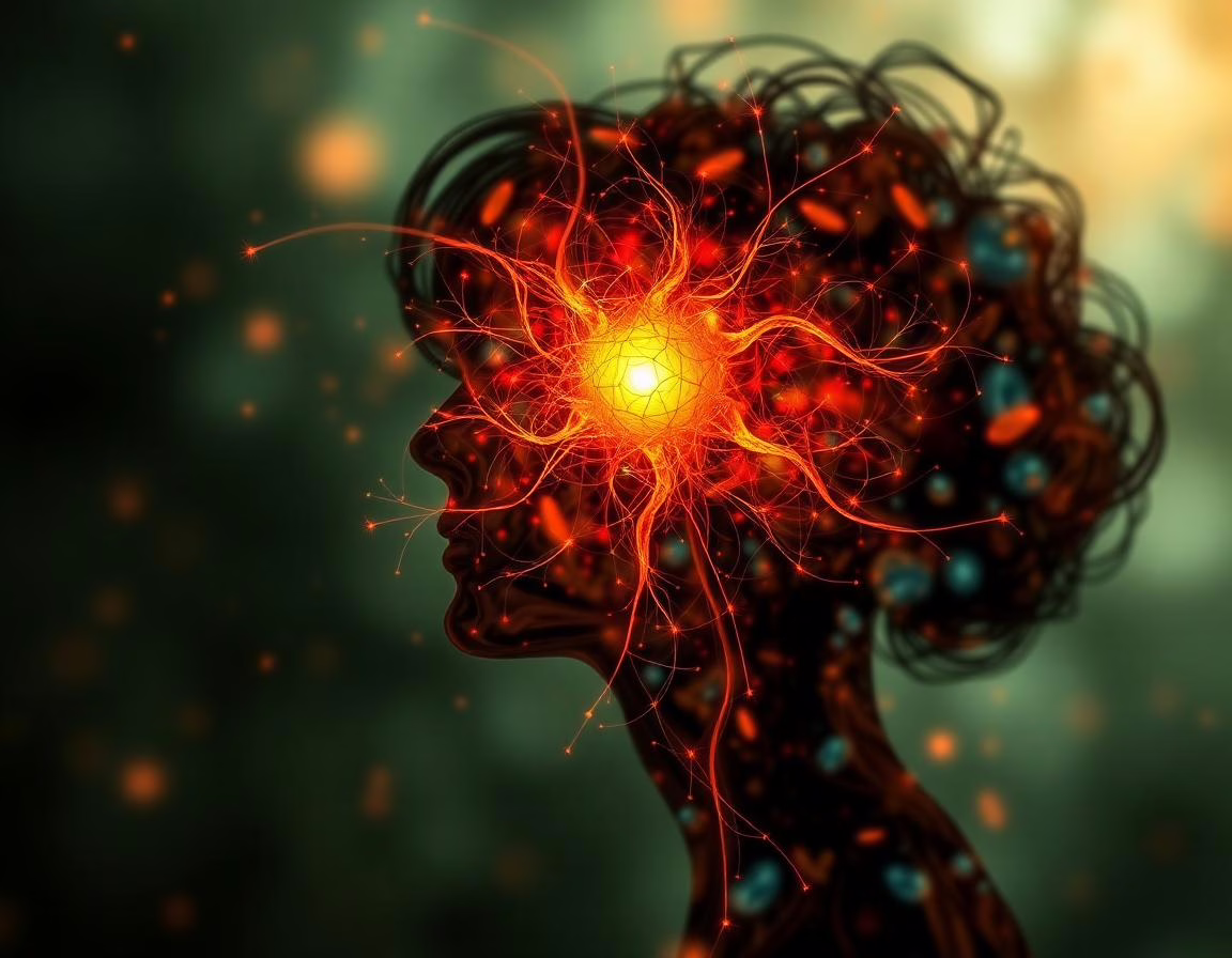
Young adults grappling with high levels of social anxiety exhibit unique patterns of increased activity and altered communication within their brain’s visual centers. Identifying these neurological changes at an early stage could pave the way for more effective detection and treatment of social anxiety before it escalates. Recent research published in a prominent journal highlights these critical findings.
Understanding Social Anxiety Disorder
Social anxiety disorder (SAD) manifests as a profound fear of social situations where one may be judged by others. This condition typically surfaces in childhood or early adulthood and can significantly disrupt personal relationships, educational pursuits, and overall quality of life. Many young individuals experience a milder form known as subclinical social anxiety, which, while not meeting the full criteria for a mental health disorder, still leads to considerable distress.
The Nature of Subclinical Social Anxiety
While many people experience shyness in new situations, subclinical social anxiety transcends typical nervousness. It involves a persistent fear of scrutiny, prompting intense physical stress responses and a strong desire to avoid social interactions altogether. By uncovering the physical underpinnings of this condition, researchers aim to validate the experiences of those who struggle with overwhelming feelings of anxiety.
Individuals with subclinical social anxiety often endure significant distress in social environments yet do not qualify for a formal diagnosis. However, the condition can still hinder daily activities and academic success. Early detection of neurological indicators may help mitigate the progression to more severe anxiety disorders.
The Research Initiative
Fangfang Huang, a researcher from Henan University of Science and Technology in China, spearheaded a study to explore the brain differences associated with early-stage social anxiety. The research team sought to understand the distinct wiring of the brains in young adults who experience heightened anxiety in social situations, aiming to pinpoint specific neurological markers linked to these anxious emotions.
The human brain comprises gray matter, which contains the cell bodies of neurons, and white matter, which forms connections between different brain regions. Investigating brain function involves examining how these areas communicate, typically assessed through blood flow measurements in the brain via magnetic resonance imaging (MRI).
Investigating Brain Activity
By observing the brain in a resting state, researchers can identify naturally communicating areas and evaluate the strength of these connections. They analyze both the overall activity levels in specific regions and the synchronized activity across different areas, determining the directionality of signals exchanged between them.
In their study, Huang and her team recruited 26 young adults with subclinical social anxiety and a comparable group of 26 healthy individuals. The participants underwent brain scans while remaining still and awake, allowing researchers to measure spontaneous brain activity over several minutes and compare the two groups.
Key Findings on Visual Processing
The researchers utilized MRI technology to track the magnetic properties of blood, measuring neural activity by observing shifts in blood oxygen levels. They focused on the amplitude of low-frequency fluctuations as an indicator of spontaneous brain activity intensity. Notably, they discovered increased activity in the left superior occipital gyrus, a region responsible for visual processing.
This heightened activity suggests that socially anxious individuals may possess an overactive visual processing system, perpetually scanning their environments for perceived social threats, such as negative facial expressions or judgmental cues.
Analyzing Functional Connectivity
The research team further examined the connectivity between the visual center and other brain regions. They assessed functional connectivity, which indicates whether two areas of the brain are active simultaneously. Findings revealed an unusually strong connection between the visual center and the right inferior frontal gyrus, a region involved in emotion regulation and social behavior monitoring.
This robust link could signify an excessive fixation on potential social threats, as the socially anxious brain remains hyper-alert, even in a resting state.
Effective Connectivity Insights
In assessing effective connectivity, the researchers tracked the directionality of signals exchanged between brain areas. They found that the visual center sent fewer signals to the postcentral gyrus, which processes physical sensations and connects emotional experiences with bodily states. Conversely, the sensory region communicated more signals back to the visual center, indicating a disruption in how anxiety is linked to visual perception.
Additionally, increased signaling from the precuneus, involved in self-reflection, to the visual center suggests that socially anxious individuals may excessively focus on their perceived flaws, reinforcing feelings of anxiety.
Structural Changes and Anxiety Correlation
The research team also investigated the gray matter volume in these brain regions to determine if structural changes contributed to the functional alterations observed. Statistical models were constructed to explore the interplay between brain structure, activity, and anxiety symptoms.
Results indicated that a reduced gray matter volume in the visual center correlated with heightened activity in that region. This overactivity subsequently predicted increased social anxiety levels among participants, illustrating a complex relationship where structural changes lead to functional hyperactivity and, ultimately, anxious experiences in social situations.
Limitations and Future Directions
While this study provides valuable insights into the neurology of social anxiety, it does have limitations. The small participant pool may restrict the generalizability of the findings. Furthermore, the homogeneity in age and education among participants raises questions about the applicability of results to a broader demographic.
Future research should include a more diverse range of participants and track individuals over extended periods to ascertain whether these brain changes persist or worsen with the progression of anxiety. Advanced imaging techniques could also enhance understanding of the physical neural pathways involved, potentially leading to targeted treatments for social anxiety.
Conclusion
This study sheds light on the neurological underpinnings of social anxiety, offering a glimpse into how altered brain activity and structure may influence this pervasive condition. As research progresses, it holds the promise of fostering innovative interventions that address early symptoms and improve the lives of those affected by social anxiety. By understanding the brain’s response to social stimuli, we may ultimately develop more effective therapeutic strategies.
- Early detection of social anxiety may prevent escalation to more severe disorders.
- Overactivity in the visual processing system is linked to heightened social anxiety.
- Strong connections between brain regions indicate an excessive focus on social threats.
- Structural changes in the brain correlate with functional hyperactivity and anxiety symptoms.
- Future studies should aim for broader participant diversity and long-term observation.
Read more → www.psypost.org