
Inflammatory arthritis poses significant challenges due to its potential for irreversible joint damage. Early detection and precise monitoring are essential for effective management. Professor Karen Knapp and her team are introducing a groundbreaking compact digital tomosynthesis (DT) system aimed at improving imaging techniques in this field. This new technology offers a low-dose, portable alternative to traditional imaging methods, providing detailed three-dimensional (3D) insights that surpass the limitations of conventional two-dimensional (2D) X-rays.
Limitations of Traditional Imaging
In the realm of inflammatory arthritis, conventional 2D X-rays are often inadequate. The overlapping of anatomical structures can obscure critical details, making it difficult to identify subtle erosions that signal disease progression. While computed tomography (CT) and magnetic resonance imaging (MRI) offer enhanced detail, they come with higher radiation exposure and are less accessible in routine clinical settings.
Digital tomosynthesis (DT) emerges as a significant advancement in musculoskeletal imaging, effectively bridging the gap between X-rays and CT. The compact DT system represents an evolution, merging the benefits of both modalities into a portable, low-dose imaging solution designed for point-of-care applications.
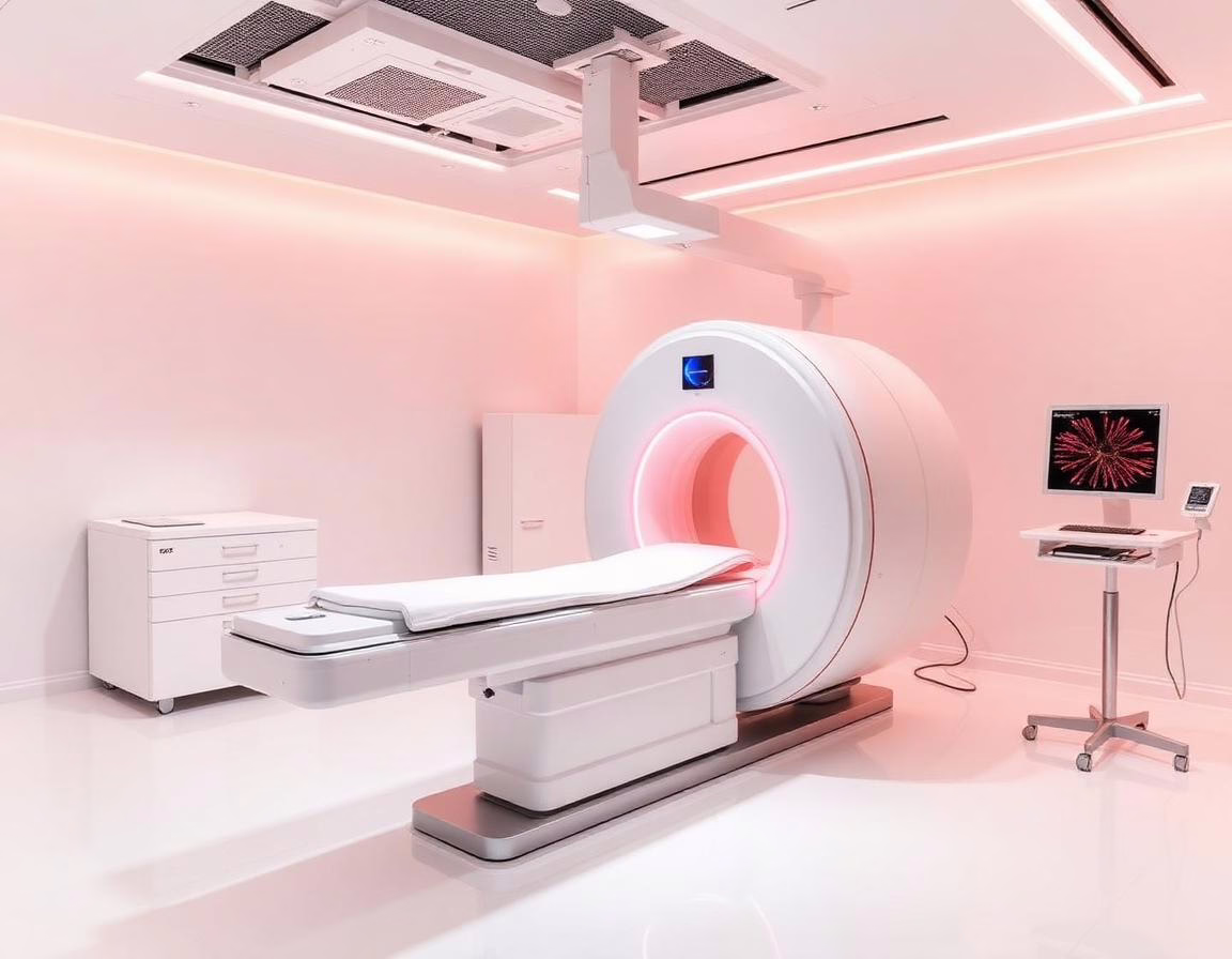
The Adaptix Ortho350: A Game Changer
The Adaptix Ortho350 device stands at the forefront of this innovation, designed for high-resolution, depth-resolved 3D imaging. By capturing multiple low-dose projections from various angles, it reconstructs these images into a detailed stack that clarifies overlapping anatomical structures. This capability allows for an unobstructed view of critical features, enhancing the detection of bony abnormalities.
Preclinical validation has already demonstrated the device’s potential. The University of Exeter has partnered with Adaptix Ltd to develop and test this compact DT scanner. Initial trials involved imaging antique human bones and animal samples, which highlighted the need for more stable imaging phantoms to simulate varying bone densities.
Advancing Towards Human Trials
The Arthritis 3D trial aims to assess the clinical efficacy of the Adaptix Ortho350 in patients with inflammatory arthritis, particularly in the hand. With rheumatoid arthritis affecting a significant portion of the adult population, timely imaging is essential to prevent severe erosive changes that can occur early in the disease.
The trial will compare the compact DT imaging results with traditional X-rays, utilizing a direct head-to-head design. A total of 30 participants will be recruited at the Royal Devon University Healthcare NHS Foundation Trust, ensuring a comprehensive evaluation of the device’s performance.
Trial Structure and Patient Involvement
Participants in the Arthritis 3D trial will undergo DT imaging within four weeks of their initial X-ray examination. This timing minimizes the risk that any observed differences in imaging results reflect disease progression rather than the imaging technique itself. Each participant will be asked to complete an acceptability questionnaire immediately after their scan.
The study’s design includes rigorous measures to eliminate bias. Radiologists will be blinded to patient identification during X-ray evaluations, and the reporting of X-ray results will occur after all scans have been completed. This structured approach aims to ensure the integrity of the trial and the validity of the outcomes.
Promising Preliminary Results
Initial findings indicate strong acceptance of the compact DT imaging among participants, coupled with excellent image quality. Enhanced visibility of complex bone structures is particularly beneficial for diagnosing subtle bone erosions and other abnormalities associated with inflammatory arthritis.
The Adaptix Ortho350 also holds promise for identifying subtle fractures, joint space irregularities, and degenerative changes, further solidifying its relevance in clinical practice.
Conclusion: A New Horizon in Imaging
The transition from preclinical validation to real-world application marks a pivotal moment in the development of compact DT technology. As a low-dose, portable solution, this imaging modality is set to revolutionize the diagnosis and monitoring of inflammatory arthritis, facilitating timely interventions that could significantly improve patient outcomes. Continued evaluation within clinical settings will further unveil the full potential of this innovative imaging approach.
- Takeaways:
- Compact DT imaging offers a low-dose, portable alternative to traditional X-rays.
- The Adaptix Ortho350 enables depth-resolved imaging, enhancing detection of bony abnormalities.
- The Arthritis 3D trial will provide critical insights into the device’s clinical applicability.
- Preliminary results indicate strong patient acceptance and excellent image quality.
- Timely imaging is crucial for preventing irreversible damage in inflammatory arthritis.
Read more → hospitalhealthcare.com