
In the intricate dance of life and death within our bodies, cancer emerges as a formidable adversary, claiming its place as thesecond leading cause of mortality worldwide. The ominous specter of metastasis, the insidious spread of cancer cells to distant organs, lurks ominously in the shadows of every diagnosis. As these malignant cells embark on their journey through the circulatory highways of the body, they seed new tumors, laying the groundwork for the tragic finale of many cancer narratives. Yet, the molecular orchestrations underlying this deadly phenomenon remain shrouded in mystery, a puzzle begging to be solved.
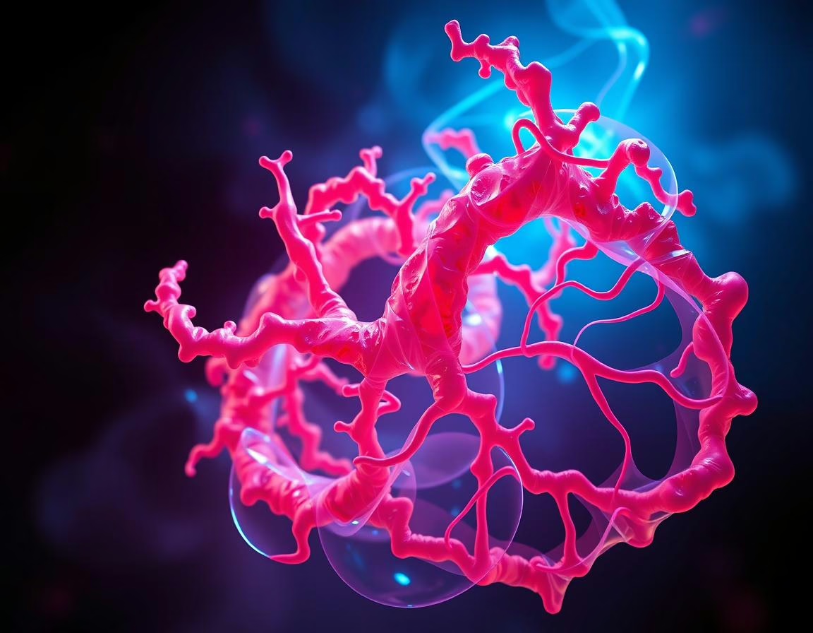
At the heart of cancer cell migration lies the cytoskeleton, a complex network of structural proteins that not only provides shape and support to the cell but also plays a pivotal role in its motility. Among these architectural elements, actin filaments (F-actin) stand out as key players in the intricate choreography of cellular movement. Their presence not only marks the path of migrating cells but also unveils secrets of cell necrosis, a harrowing consequence of membrane breakdown.
Diving into the Depths of Cellular Dynamics
Traditionally, our explorations of F-actin dynamics in cancer cells have been confined to the flat landscapes of 2D imaging, offering only a fragmented view of the cellular terrain that unfolds in three dimensions. Tumors, with their labyrinthine structures and diverse cell populations, demand a more comprehensive approach to unravel the mysteries of metastasis. To truly grasp the nuances of cytoskeletal involvement in cancer spread, we must embrace imaging techniques that can capture the volumetric intricacies of cellular behavior.
A recent study conducted at the Vienna University of Technology, Austria, ventured into this uncharted territory using light sheet fluorescence microscopy (LSFM) to illuminate the cytoskeletal alterations within spheroids composed of colorectal cancer (CRC) and non-small cell lung carcinoma (NSCLC) cells in a 3D setting. By deconvoluting 2D data with NeuroDeblur and reconstructing it into a 3D landscape with Thermo Scientific Amira Software, the researchers uncovered a rich tapestry of internal and external tissue structures, shedding light on the hidden realms of cellular dynamics.
Peering into the Heart of Cancer Cells
Employing a laser to induce fluorescence across successive cross-sections of a sample, light sheet fluorescence microscopy enables precise optical sectioning of the specimen, paving the way for the creation of a 3D representation that captures the essence of cellular life in its entirety. In the hands of Prado-López and colleagues, this cutting-edge technology revealed the inner workings of CRC and NSCLC cell spheroids, exposing internal structures suffused with autofluorescence that facilitated differentiation and segmentation. Here, amidst the intricate cellular tapestries, F-actin structures emerged as beacons of insight, their intensity quantified in individual cells to unveil hidden truths.
Through meticulous segmentation and analysis using Amira Software, the researchers traced the intricate dance of F-actin intensity across different cellular states, from proliferation to senescence and ultimately to necrosis. This journey into the heart of cancer cells unraveled the intimate connection between F-actin polymerization, cell stiffening, and enhanced cell mobility, providing a window into the molecular mechanisms that drive cancer metastasis.
Charting a New Course towards Therapeutic Discoveries
As we stand at the threshold of a new era in cancer research, armed with the power of 3D fluorescence microscopy to unravel the enigmatic complexities of metastasis, we are poised to make significant strides towards unraveling the mysteries of this deadly process. The ability to visualize cancer cell mobility at a molecular level opens up avenues for targeted interventions and novel treatment modalities, promising hope to countless individuals grappling with the specter of metastatic cancer.
In conclusion, the journey into the molecular realm of cancer metastasis, guided by the illuminating beacon of 3D fluorescence microscopy, holds the promise of transformative discoveries that could reshape the landscape of cancer therapy. By peering into the intricate tapestry of cellular dynamics, we inch closer towards unlocking the secrets of metastasis and offering new hope to those in the relentless grip of this insidious disease.
Takeaways:
– 3D fluorescence microscopy unveils the hidden intricacies of cancer metastasis, shedding light on the molecular basis of this deadly process.
– Actin filaments (F-actin) emerge as key players in cancer cell migration, offering insights into cell motility, necrosis, and potential therapeutic targets.
– Light sheet fluorescence microscopy enables precise 3D visualization of cellular dynamics, providing a comprehensive view of tumor structures and cytoskeletal alterations.
– Segmentation and quantitative analysis of F-actin intensity reveal the intimate connection between cell stiffness, mobility, and the progression of cancer metastasis.
– The insights gained from 3D fluorescence microscopy pave the way for targeted interventions and novel treatment strategies in the battle against metastatic cancer.
Read more on news-medical.net